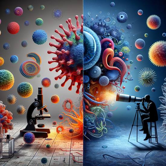
In the realm of scientific inquiry and medical advancement, the ability to visualize the invisible has always been a game-changer. This is especially true in virology, where understanding the intricate details of viruses is crucial for developing treatments and vaccines. But how can scientists take pictures of entities as minuscule and elusive as viruses? The answer lies in groundbreaking technology known as Cryo-Electron Microscopy (cryo-EM), a technique that has revolutionized structural biology and virus research. At Shuimu Biosciences, we leverage this Nobel Prize-winning technology, combined with artificial intelligence (AI), to push the boundaries of what’s possible in drug discovery and development.

Cryo-EM services have emerged as a cornerstone in the field of structural biology, allowing scientists to visualize biological molecules in their native state without the need for dyes or crystallization. This is particularly beneficial for studying viruses, which are too small for traditional light microscopy and too delicate for conventional electron microscopy. Shuimu Biosciences, a pioneer in “AI+ Cryo-EM” driven pharmaceutical research acceleration, operates the largest commercial cryo-EM technology service platform globally. Our expertise in cryo-EM analysis has facilitated groundbreaking insights into viral structures, mechanisms of infection, and paths to neutralization.
Cryo-EM involves flash-freezing biological samples to preserve their natural structure and then bombarding them with electrons to capture images. These images are then reconstructed into detailed 3D models using advanced computational algorithms. Shuimu Biosciences has enhanced this process through the development of our proprietary SMART software and computing platform, which integrates AI to streamline data collection, analysis, and modeling. This not only accelerates structural analysis but also improves the resolution and accuracy of the resulting images.
Cryo-EM’s Impact on Viral Research
The ability to take high-resolution pictures of viruses using cryo-EM services has had a profound impact on virology. It enables researchers to understand the precise mechanisms by which viruses invade host cells, how they replicate, and how they evade the immune system. This knowledge is crucial for the design of effective antiviral drugs and vaccines. Shuimu Biosciences’ cryo-EM analysis has contributed to significant advancements in this field, including the characterization of novel viral targets and the optimization of antiviral compounds.
Shuimu Biosciences: At the Forefront of Cryo-EM Services
At Shuimu Biosciences, we are committed to advancing the frontiers of life sciences through our comprehensive cryo-EM services. Our state-of-the-art cryo-EM facilities, including eight 300kv high-end microscopes, serve over 500 innovative pharmaceutical companies and research institutions worldwide. Our efforts in cryo-EM analysis not only support the global scientific community but also accelerate the path to new treatments for diseases caused by viruses.
In conclusion, taking pictures of viruses is not only possible but has become a fundamental aspect of modern virology, thanks to cryo-EM. Shuimu Biosciences is proud to be a leader in this field, offering unparalleled cryo-EM services and analysis to aid in the fight against viral diseases. By harnessing the power of cryo-EM and AI, we are opening new doors in drug discovery and development, bringing hope to millions affected by viral infections around the globe.
For more information on how our cryo-EM services can support your research and development projects, visit Shuimu Biosciences today. Together, we can unlock the mysteries of viruses and pave the way for a healthier future.
